Training for operating a scanning electron microscope(SEM)
A downloadable tool for Windows
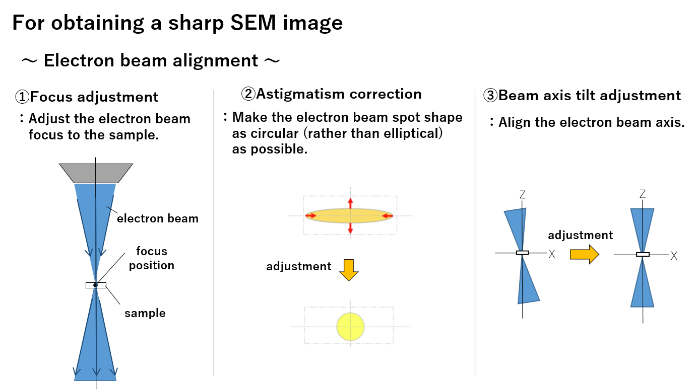
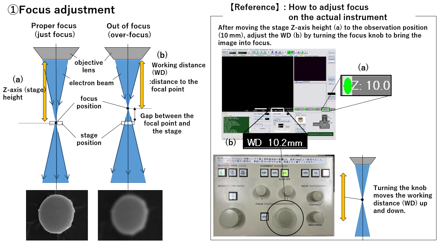
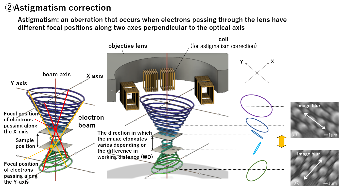
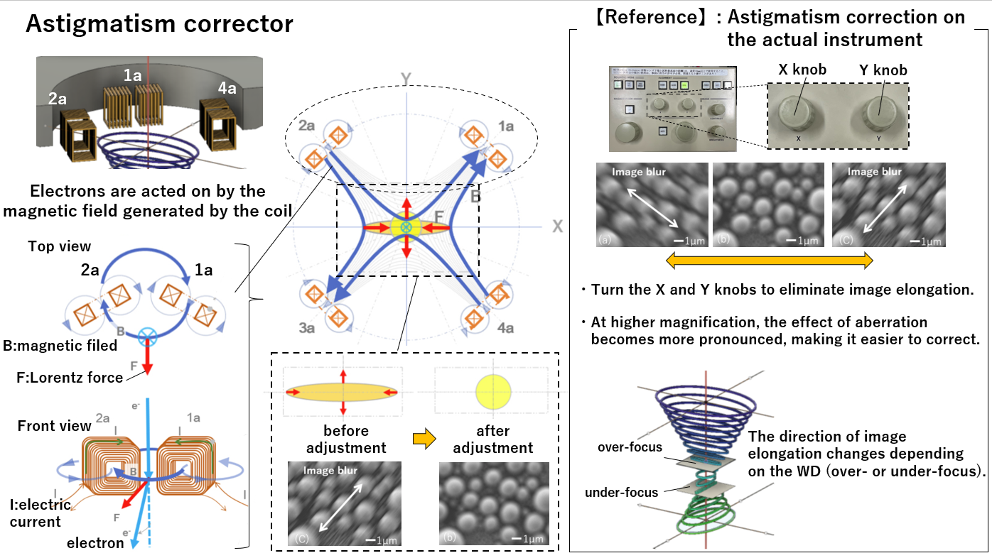
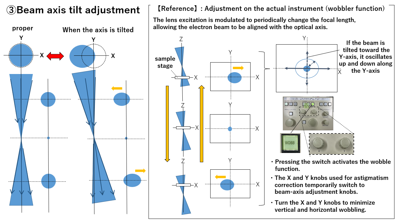
Scanning electron microscopes(SEM) irradiate the sample with an electron beam to obtain magnified images of micro‑scale structures. However, the beam’s focal distance, astigmatism, and axial misalignment directly affect the direction and degree of image blurring. Although automation such as auto‑adjustment has advanced in recent years, observing nanometer‑scale features still requires a high level of operational skill, and establishing an efficient training method remains a challenge.
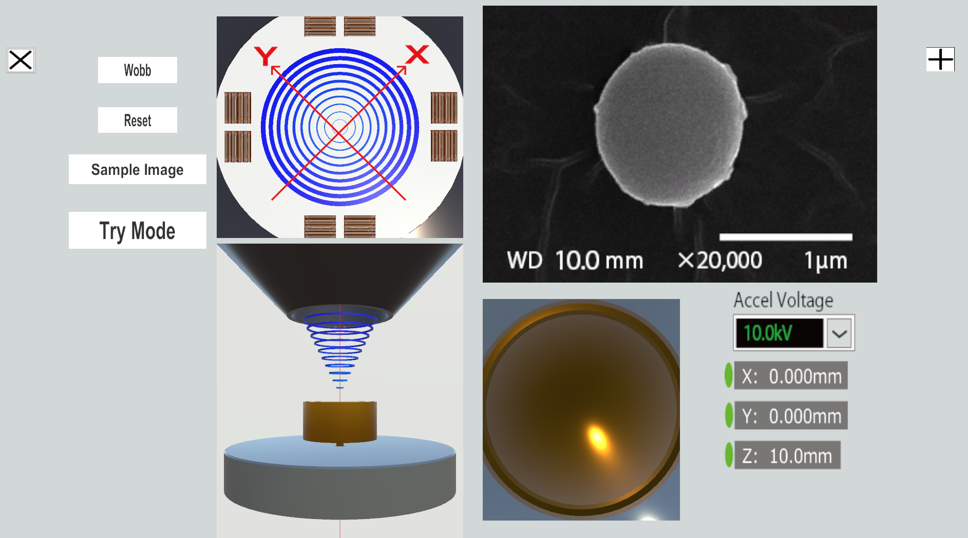
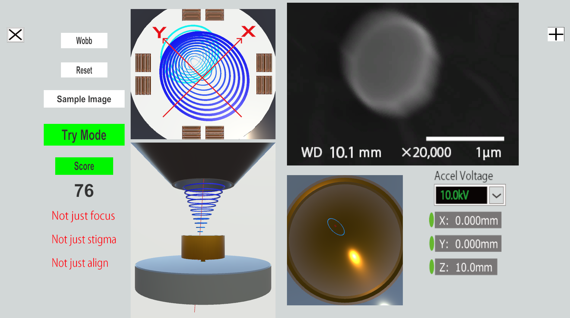
This application is designed to help users understand the relationship between the electron beam and the observed image, and to support skill acquisition through practice in adjusting the electron beam.
The simulated images in the application are based on actual SEM images of approximately 1 µm polystyrene particles observed at 20,000× magnification. Using these images, a model was created that reproduces the direction and degree of blurring, and the displayed image changes according to the user’s operations.
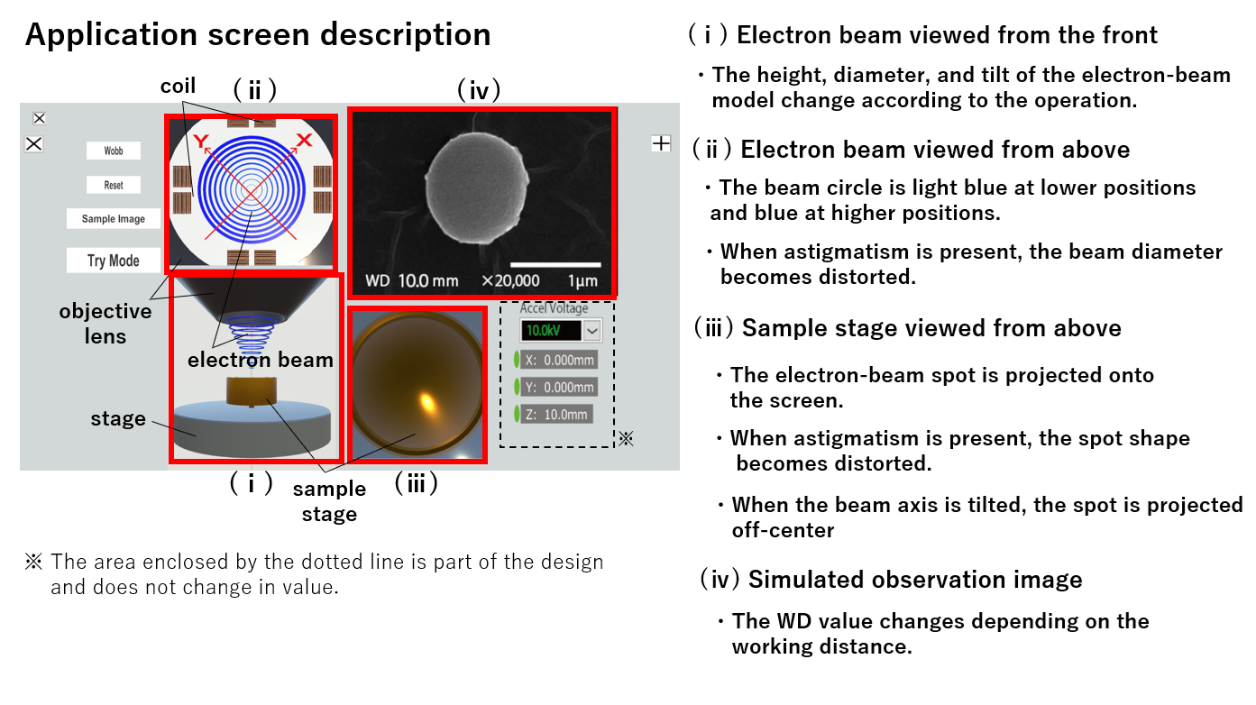
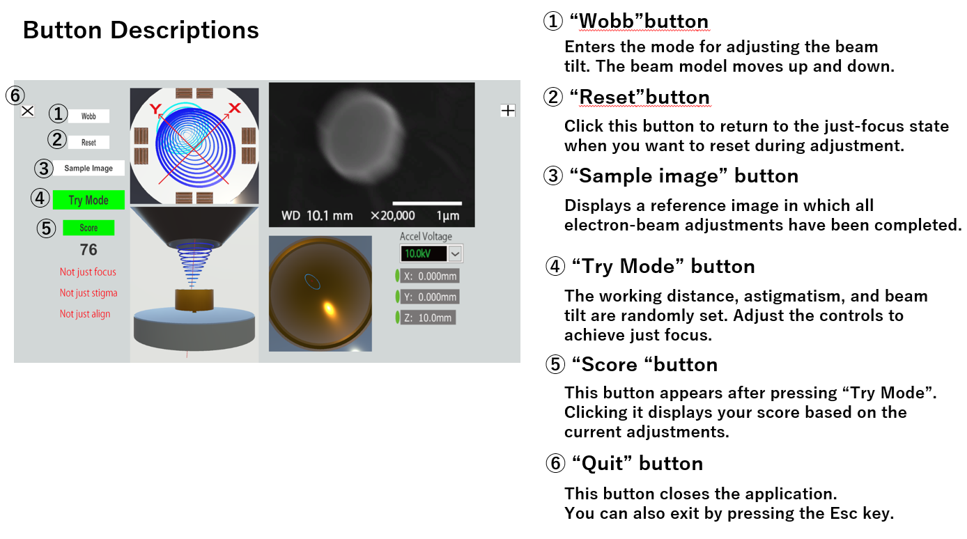
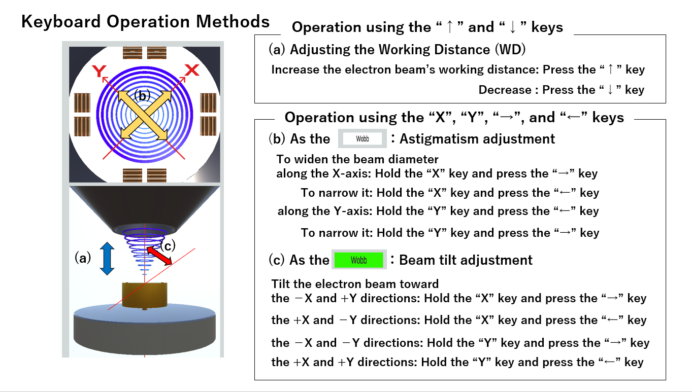
Instructions for operation are provided in the attached images.
Download
Install instructions
Please extract the downloaded ZIP file.
After extracting, double-click “My project.exe” inside the folder to launch the application.
No installation is required.
Please do not change the file structure inside the folder.